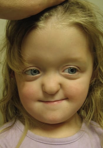
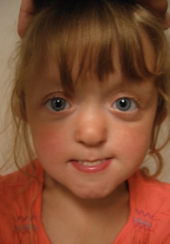
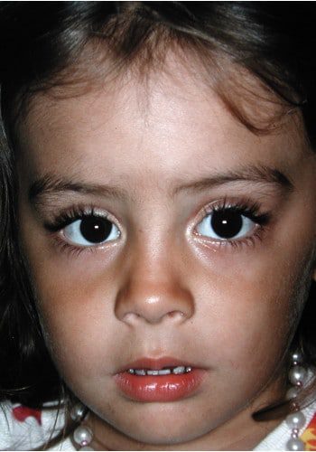
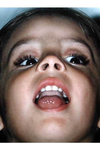
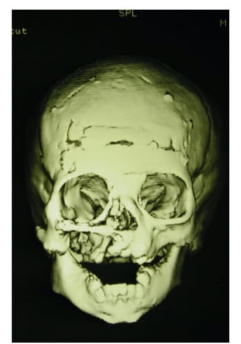
pwsbuilder

A young boy with an open mouth and two different eyes.

Craniofacial

Case ID: 4253

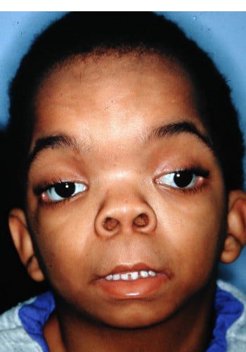
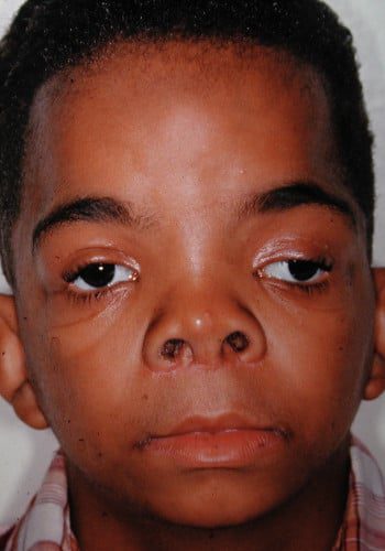
Front: Crouzon Syndrome with hypertelorism. Post-op correction with cranial bone graft to nose.
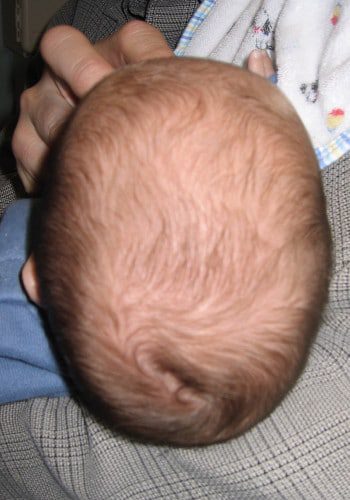
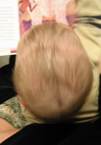
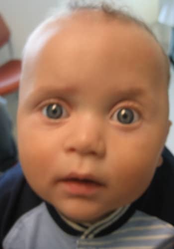
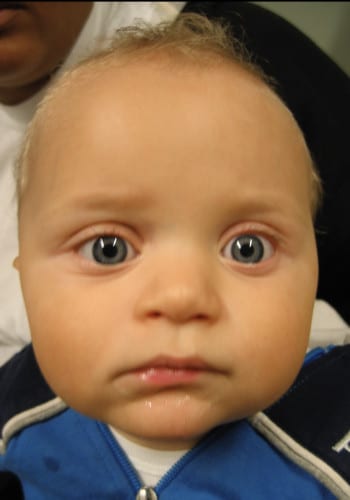
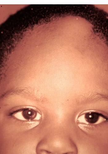
A before and after picture of a baby 's head.

Craniosynostosis

Case ID: 4386

Front: Pre and 2 months post-op correction of sagittal synostosis.

A baby and a young girl with very large eyes.

Craniosynostosis

Case ID: 4434

Front: Metopic Synostosis pre and post-op.

Front: Shape of forehead pre and post-op.
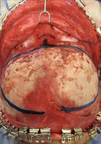
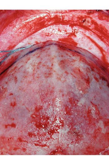
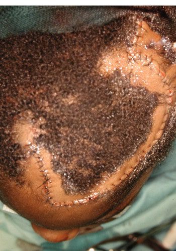
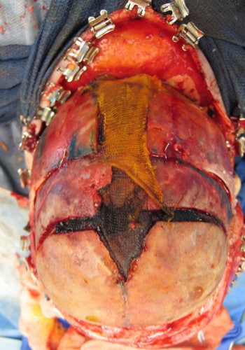
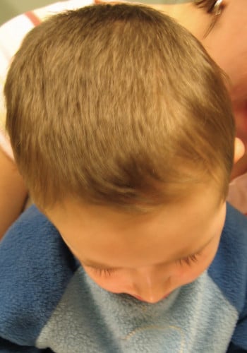
A child with an ear infection and a head of hair.

Craniofacial Tumor

Case ID: 4827

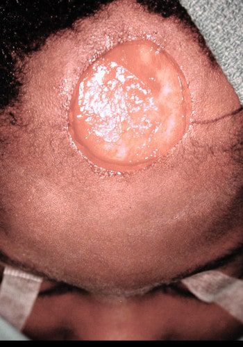
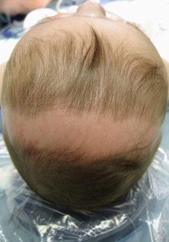
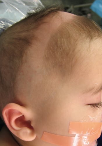
Front: Young boy with growing fibroma of scalp and frontal bone. Image of excision.
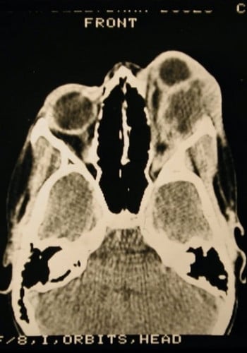
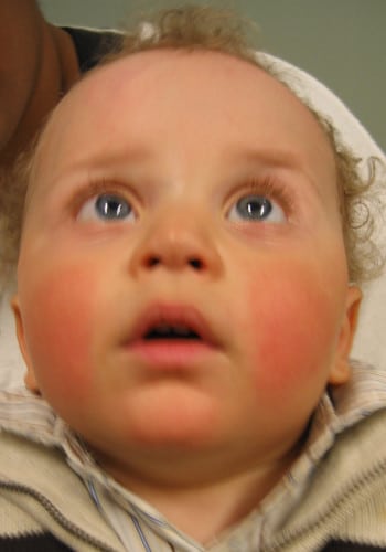
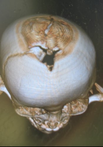
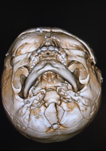
Front: Intra-op closure with rotation flap and long term result.

A baby with an apple in its mouth and a radiograph of the same image.

Craniofacial Tumor

Case ID: 4942

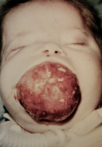
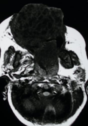
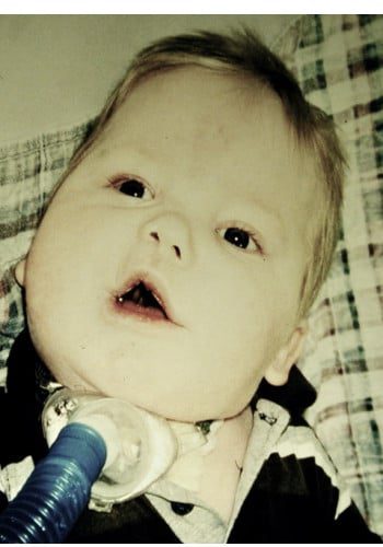
Front: Infant with massive teratoma obstructing airway. CT of tumor.

Front: Complete resection of tumor in one procedure with neurosurgery; Reconstruction of maxilla with cranial bone graft and repair of palatal cleft defect.

A baby with a bandage on his head and a man with a bandaged forehead.

Craniosynostosis

Case ID: 4515

Front: Sagittal Craniosynostosis pre op

Front: Intra-op view and post-op photo.
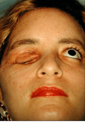
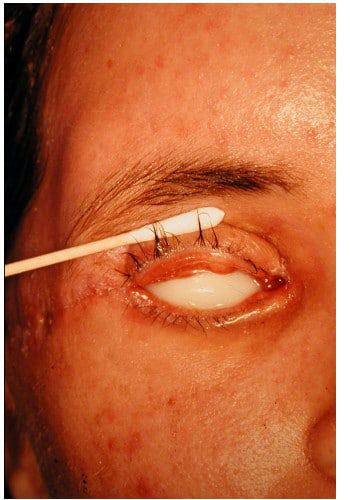
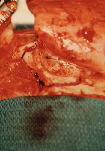
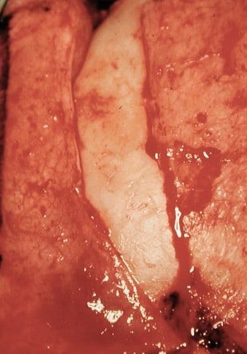
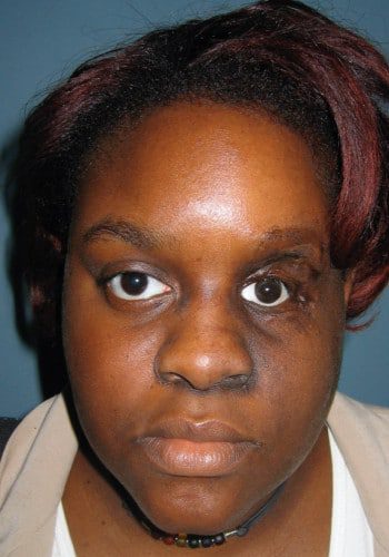
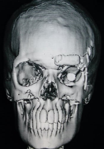
A woman with an eye injury and a picture of the same person

Craniofacial Tumor

Case ID: 5023

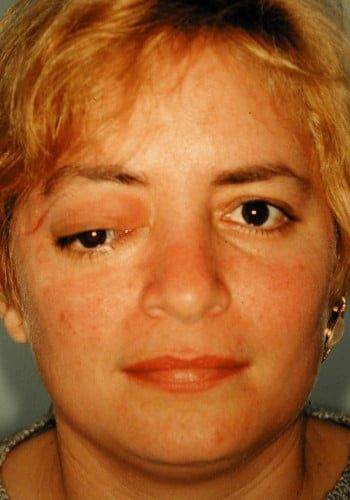
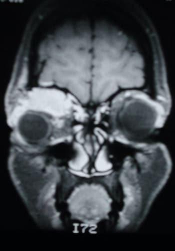
Front: Young woman with right orbital Lacrimal Adenocarcinoma, recurrent with metastasis to frontal lobe of brain.

Front: After radical exenteration of tumor and involved dura; preservation of uninvolved eyelids; no recurrence in 25+ years.

A young boy with an eye injury and an x-ray of the same condition.

Craniofacial Tumor

Case ID: 5029

Front: 10 year old girl with Rhabdomyosarcoma of left orbit after radiation and chemotherapy.

Front: Intra-op views of orbital bone excision and preparation of periosteal-temporal muscle flap to insert into exenterated orbit.

Front: Post-op 12 years; no recurrence of tumor; bony reconstruction of orbit and insertion of stable prosthetic globe and preservation of eyelids.

Two pictures of a young girl with and without makeup.

Craniosynostosis

Case ID: 4586

Front: Young girl with Crouzon Syndrome pre and post-op Le Fort III

A split photo of two babies with one looking surprised.

Craniosynostosis

Case ID: 4636

Front: Left unilateral coronal craniosynostosis pre and post-op.

Front: Intra-op view of retrusion left fronto-orbit region and post-op correction.

Front: Pre-op CT with absent left coronal suture and retrusion fronto-orbit. Post-op CT.