pwsbuilder

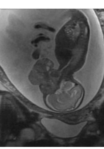
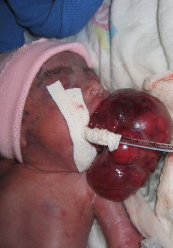
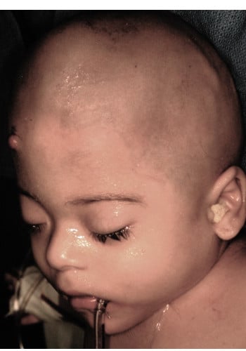
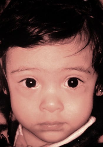
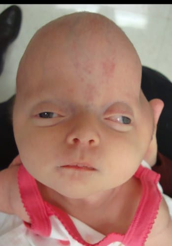
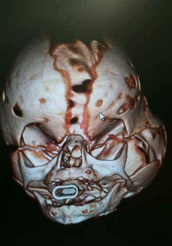
Craniofacial Tumor
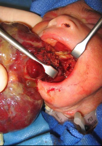
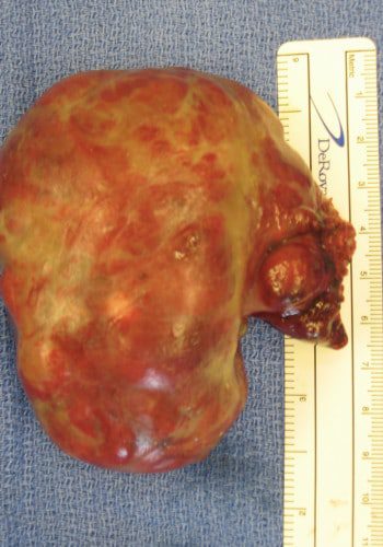
Case ID: 4619
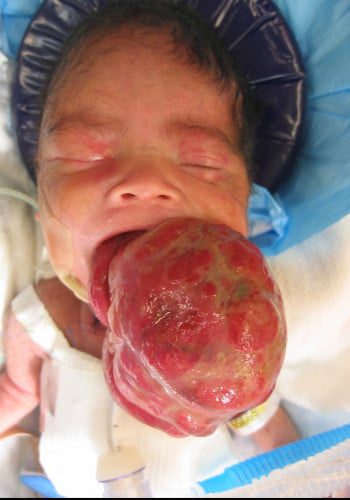
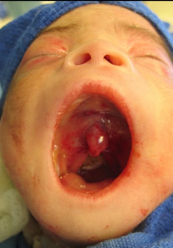
Front: Newborn with giant teratoma after emergency tracheostomy. Post op resection with neurosurgery and simultaneous repair of cleft palate.

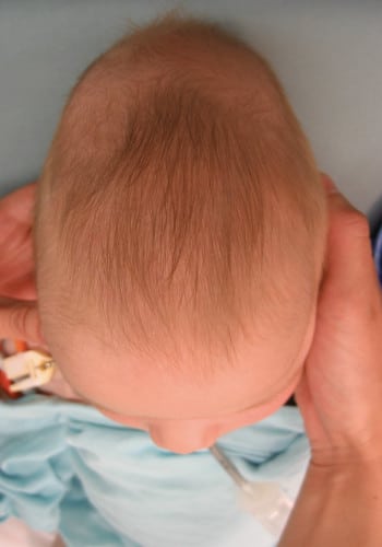
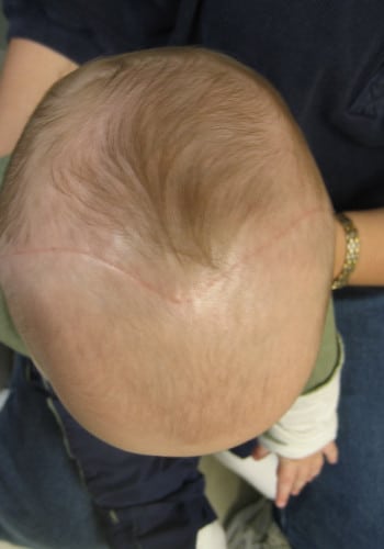
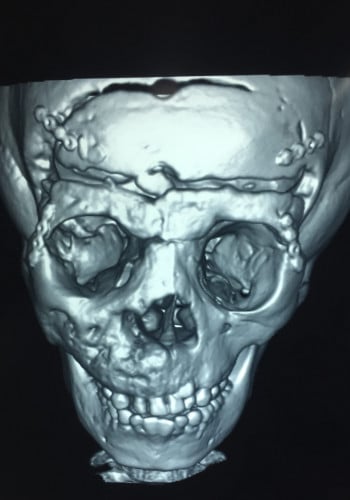
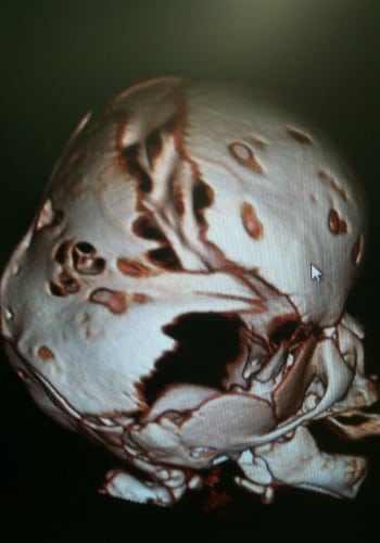
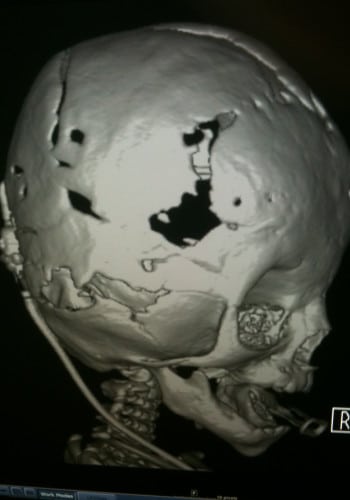
Craniosynostosis
Case ID: 3961
Front: Saethre-Chotzen Syndrome (Bicoronal synostosis)
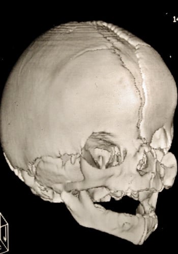
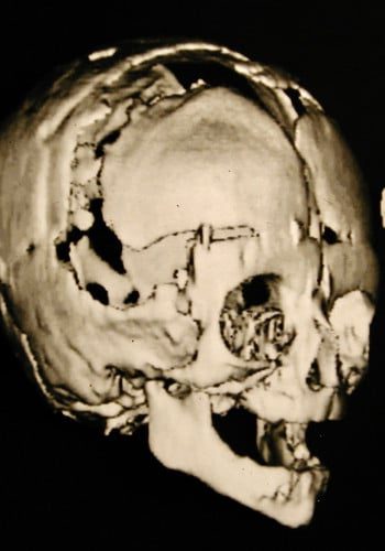
Front: Pre and post craniotomies and front-orbital advancement.


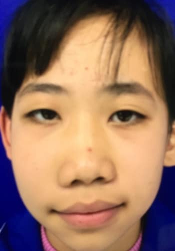
Craniofacial Tumors
Case ID: 4649
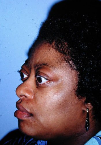
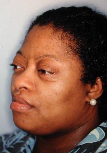
Front: Patient with Graves Disease, exophthalmos and eyelid retraction pre and post op.

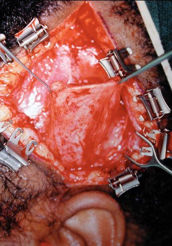

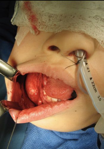
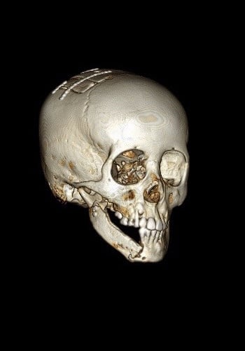
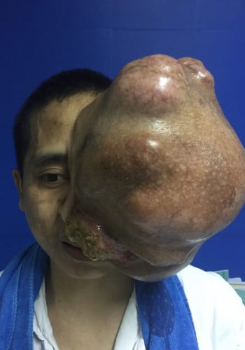
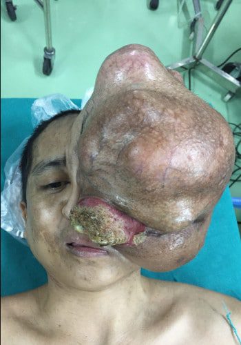
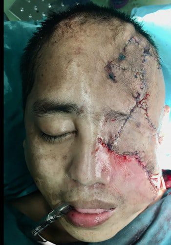
Craniofacial Tumor
Case ID: 4686

Front: 32 y.o. woman with left temporal Fibrous Dysplasia, pre and post-op radical resection and bone grafting.
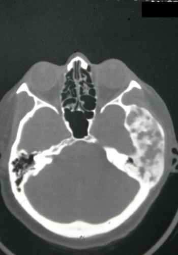
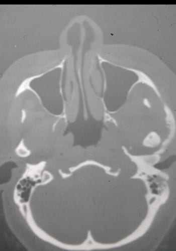

Craniofacial Tumor
Case ID: 4740
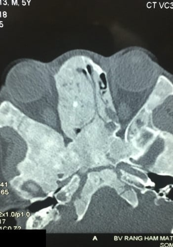
Front: Young child with Fibrous Dysplasia of right orbital-ethmoid region.


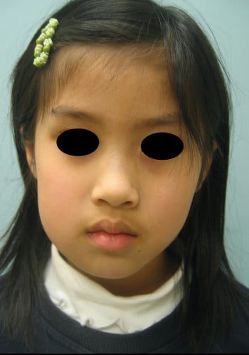
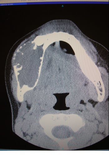
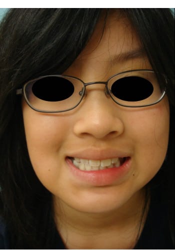
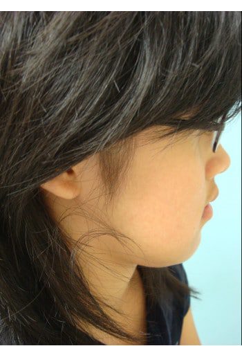
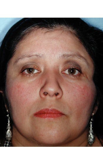
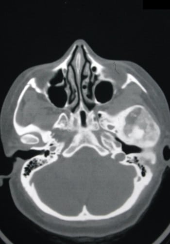
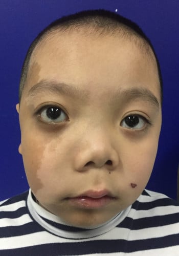
Craniofacial Tumor
Case ID: 4750
Front: Young boy with right maxillary Fibrous Dysplasia and orbital dystopia.


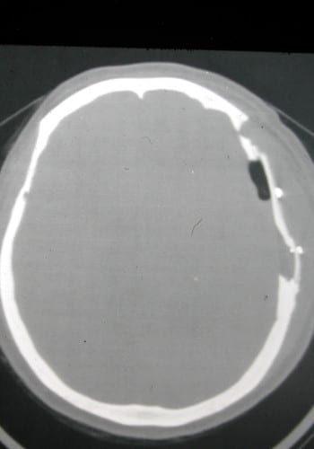
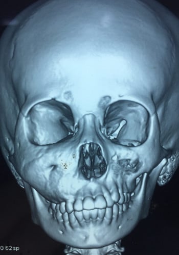
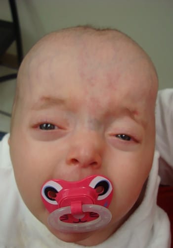
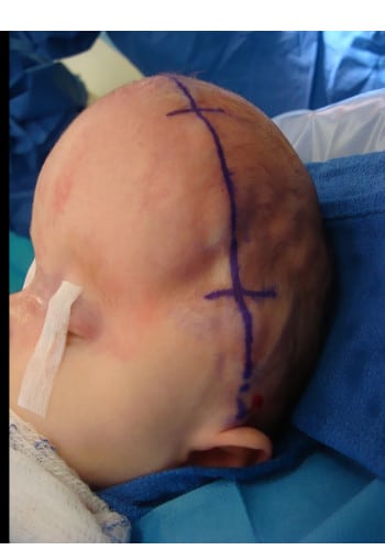
Craniosynostosis
Case ID: 4188
Front: Pre-op 3 month old with Crouzon Syndrome and most sutures fused. Note left strabismus and exorbitism.
Front: Post op images after (one operation) multiple craniotomies, front-orbital advancement, and post-op distraction osteogenesis.
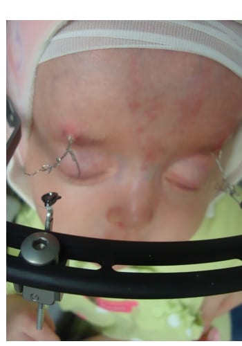
Front: Intra-operative view and post-op 4 week distraction (device invented by Dr. McKinnon).
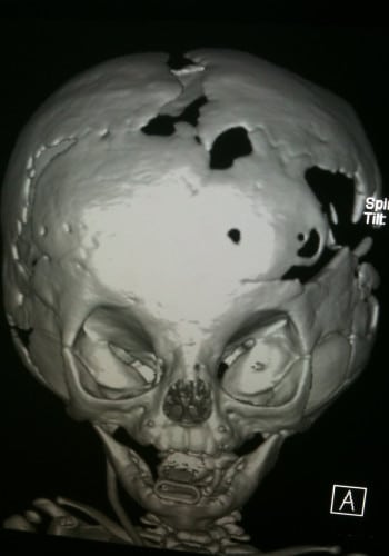
Front: Pre and post-op CT images showing vast increase in cranial volume.